Describe the Uses of Dental Imaging Chapter 38
They can be divided into periapical bitewing and occlusal projections. Many people think that dental treatment is a painful procedure.
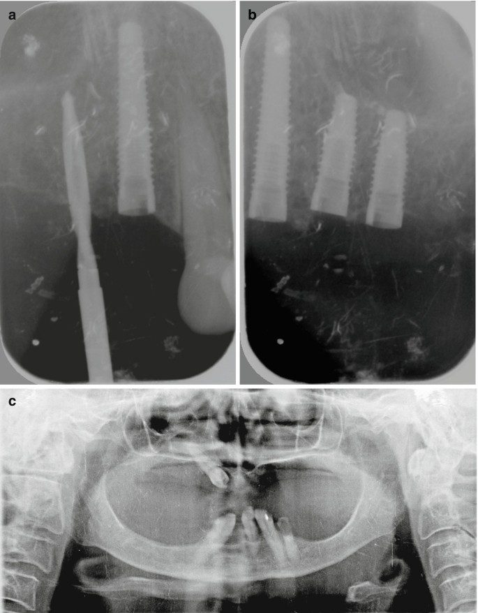
Technical Errors And Artefacts In Dental Radiography Springerlink
List protective measures and methods used to reduce the risk principle in radiation exposure.
. Study Chapter 38 flashcards. Describe the use of dental imaging. Process of making radiographys of the teeth and adjacent structures exposure to radiographs.
DA130 Dental Terminology Chapter 38 2docx Kilovoltage. If you have any remaining questions or concerns about the X-ray process please give our office a call at 720 409-0008. Identify bone loss in the early stages.
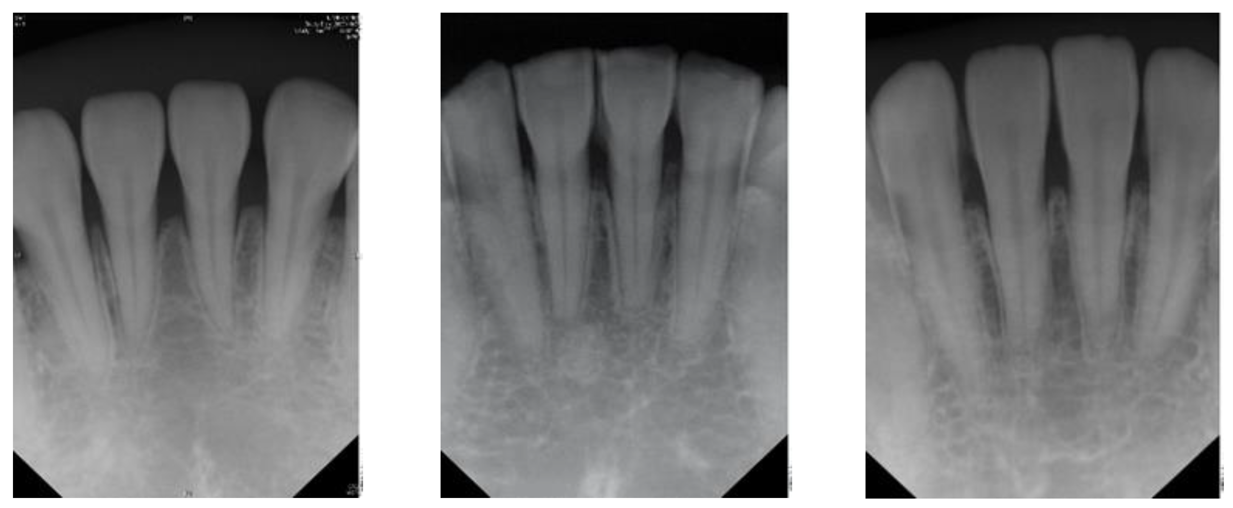
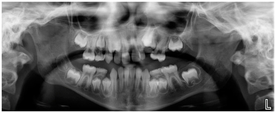
Dental radiology includes the periapical film PAX to visualize periapical pathology bitewing films to identify occlusal and interpromimal dental caries occlusal films most commonly to identify submandibular sialolithiasis the panorex panoramic radiograph or orthopantomogram is a two-dimensional view of the bones and dentition of the upper and. Intraoral bi-dimensional radiographs compose an essential part of dental imaging. The evolution of 3-dimensional imaging.
You also learned aboutthe XCP which stabilizes a receptor when taking an image. In this chapter you learned aboutthe differentparts of an Xray unit and whatthey do to produce diagnostic images with good contrastand density. A unit of measurement used to describe the intensity of an electrical current.
Foundatins of Radiography Radiographic Equipment and Radiation Safety. Overall darkness or blackness of a radiograph. 38 Quantity of x-ray beam define 39 Volt V define 40 Voltage.
A unit of measurement used to describe the intensity of an electrical current. Item placed over patient to protect the reproductive and blood forming tissues from scatter radiation Magnificationproportional enlargement of a radiographic image Master switchcomponents of. Chapter 38 MDA Flashcards.
The uses of dental images include the detection of abnormalities in surrounding hard and soft tissues Exposure to Study Resources. Willis July 21 2017 Short- Answer Questions 1. Identify the unit of measurement used to describe the amount of electric current flowing through the x-ray tube.
Dental X-ray tube acts as a self-rectifier in that it changes AC to DC while producing x-rays this ensures that the current is always flowing in the same direction from cathode to anode What circuits are involves with x-rays. You will learn more aboutthe use of the XCP in Chapter 3. Filmless method of capturing an image and displaying it by using an image sensor and electronic signal and a computer to process.
Bringing awareness among individuals about dental treatment and educating the individual about dental treatment that it will help in maintaining good oral hygiene which is needful for maintaining good health creates a positive attitude in the individual. DENT 1050 Chapter 3 Flashcards Preview Dental Radiography DENT 1050 Chapter 3 Flashcards Flashcards in DENT 1050 Chapter 3 Deck. The three-dimensional printing has been used since very long ago in the field of medicine as well as in dentistry.
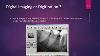
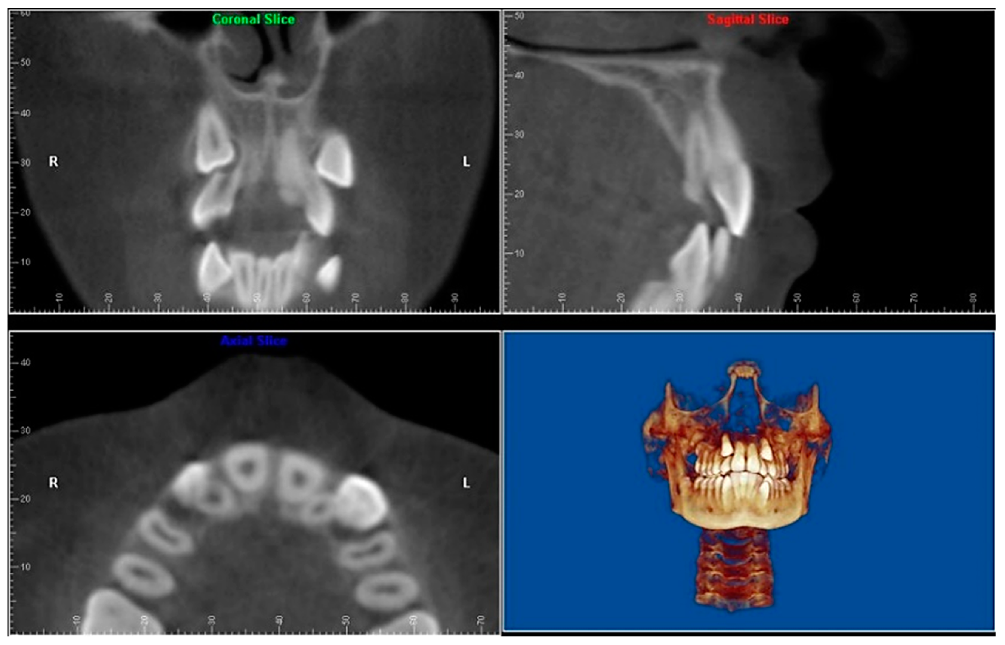
Medical imaging is the process of creating visual pictures of the inside of the human body for diagnostic and treatment purposes. BirdRobinson Modern Dental Assisting 10th edition Chapter 38. Describe the purpose and uses of cone beam computed tomography.
The uses of dental imaging are checking patients oral health and making it clearer to diagnose certain teeth problems such as cavities. Describe the discovery of x-radiation. Stage that has not developed yet Lead apron.
View Test Bank - Chapter 38docx from DEH DES at Miami Dade College Miami. Highest voltage of radiograph tube used during a radiograph exposure Latent. Evaluate growth and development.
Identify dental radiographic techniques and list the uses of radiographs in dentistry. The ring indicates the boundary of the receptor. Katherine Moran Radiology Mrs.
32 rows The process of of recording images of the teeth and adjacent structures by exposure to x-radiation. Uses of digital imaging-To detect lesions diseases and conditions of teeth and surrounding structures-Confirm or classify suspected disease-Provide information during dental procedures root canal therapy instrumentation and surgical placement of. Is the blurred or indistinct area that surrounds an image.
Provide information during dental procedures such as root canal therapy Document a patients condition at a specific time. Describe the uses of dental imaging. Describe the interactions of dental x-rays with matter and action on tissues and cells.
A filmless method of capturing an image and displaying it by using an image receptor an electronic signal and a computer to process and store the image. We use dental X-rays as a diagnostic tool to help us pinpoint any problems with your teeth and gums that require attention. Locate abnormalities in surrounding hard and soft tissues.
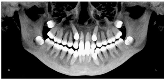
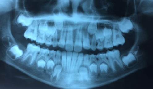
Another widely used dental image is the panoramic exam which is based on an extraoral technique and produces a single tomographic image of the facial structures including teeth mandibles and adjacent structures. Is one one-thousandth 11000 of an ampere. One one-thousandth of an ampere.
The cone beam computed tomography is used to view the area of the head and neck in three dimensions and it is used to find the exact placement of implants the buccallingual position of impacted teeth to be removed and determination of the exact location of the mandibular nerve before surgery is done. Differentiate between the different types of radiation. Detect dental caries in the early stages.
Dentistry Journal Free Full Text Cone Beam Computed Tomography In Orthodontics Html

Basic Terminology Of Dental Radiography Video Lesson Transcript Study Com
Sensors Free Full Text Detection Of Dental Apical Lesions Using Cnns On Periapical Radiograph Html
Sensors Free Full Text Detection Of Dental Apical Lesions Using Cnns On Periapical Radiograph Html

Sample Panoramic Dental Radiographs And Annotated Pbl Lesions Top Download Scientific Diagram
Clinical Management Of Hypoplasic Amelogenesis Imperfecta A Challenge For Multidisciplinary Team A Case Report
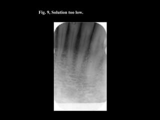
Lecture 7 Dental X Ray Film Processing And Processing Errors Lecture

Bone Quality Of The Dento Maxillofacial Complex And Osteoporosis Opportunistic Radiographic Interpretation Intechopen

Pdf History Of Dental Radiography Evolution Of 2d And 3d Imaging Modalities
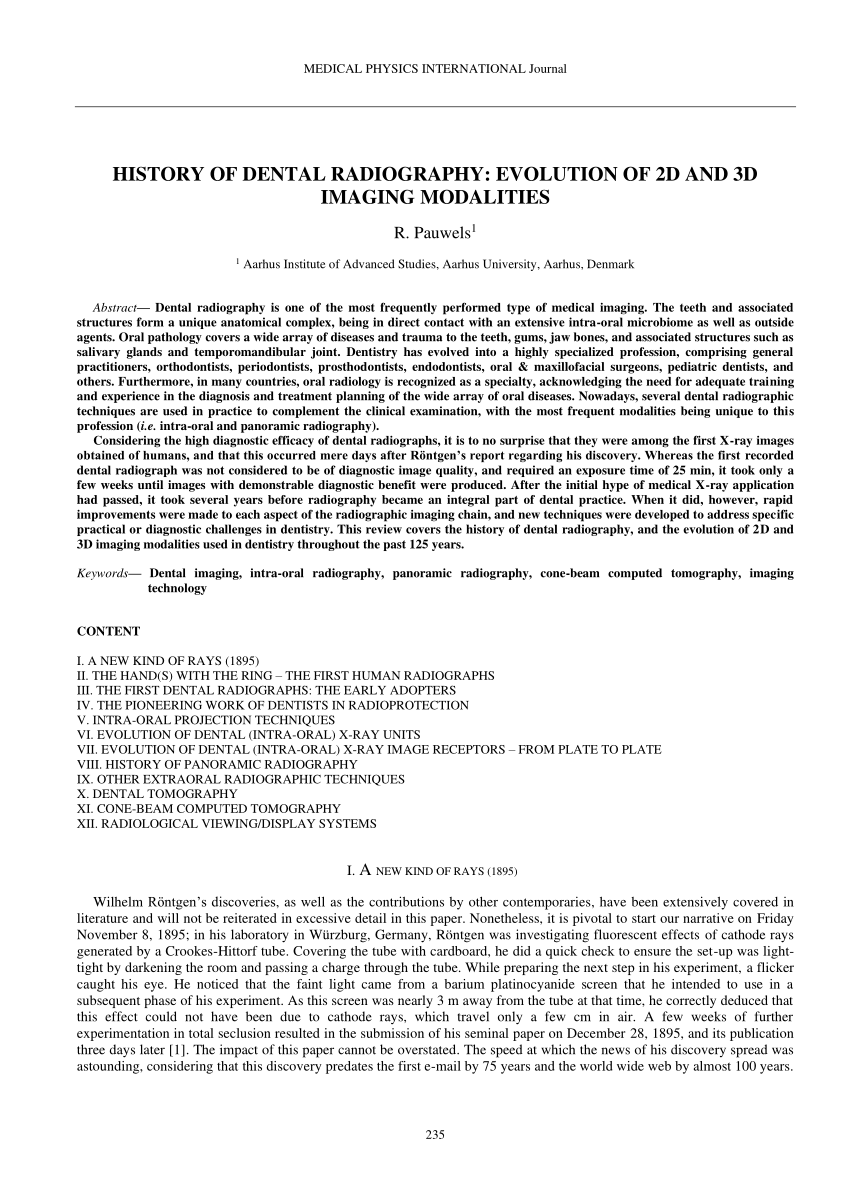
Pdf History Of Dental Radiography Evolution Of 2d And 3d Imaging Modalities

Pin On Work
Dentistry Journal Free Full Text Cone Beam Computed Tomography In Orthodontics Html
Digital Imaging In Dentistry
Digital Imaging In Dentistry

Radiographs In Periodontal Disease Diagnosis And Management Corbet 2009 Australian Dental Journal Wiley Online Library

Pin On Orthodontic Instrument

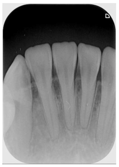
Periapical Radiography Dental Life Radiography Intraoral
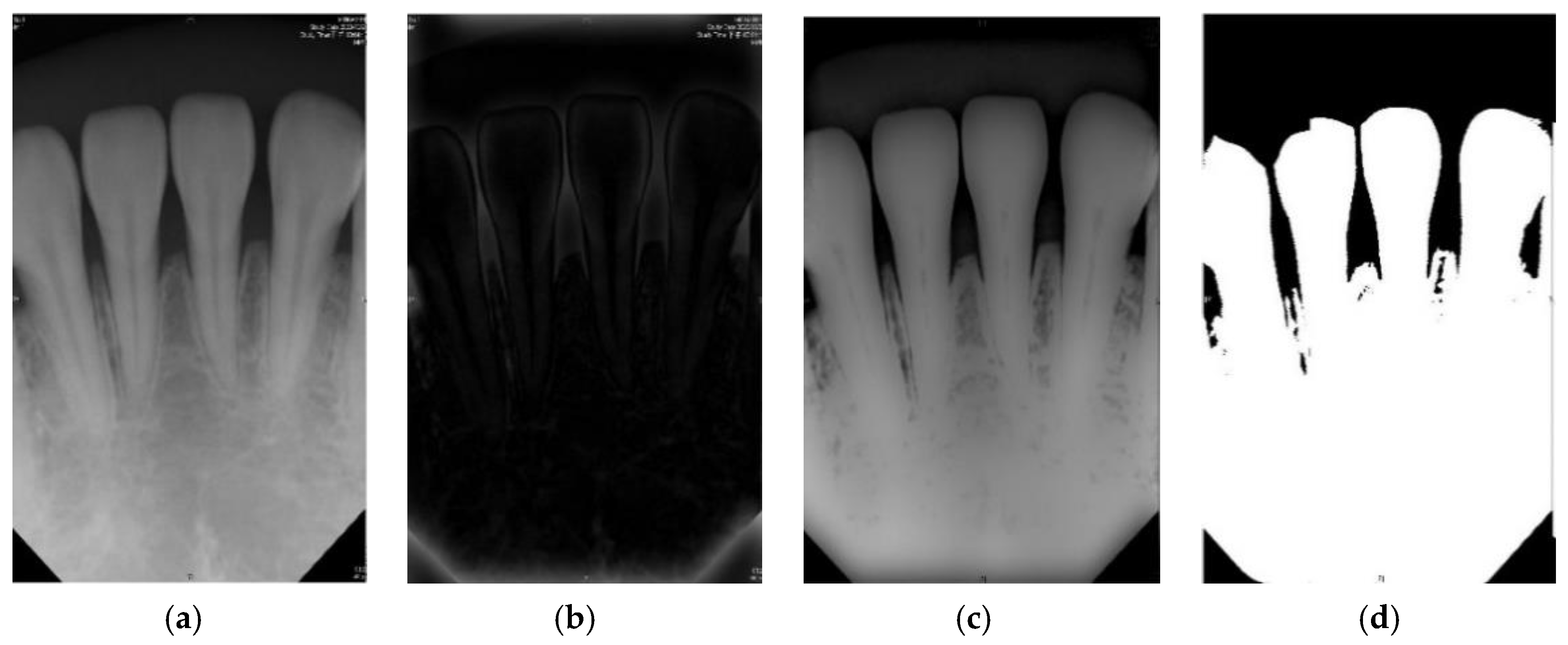
Sensors Free Full Text Detection Of Dental Apical Lesions Using Cnns On Periapical Radiograph Html
Dentistry Journal Free Full Text Cone Beam Computed Tomography In Orthodontics Html


Comments
Post a Comment